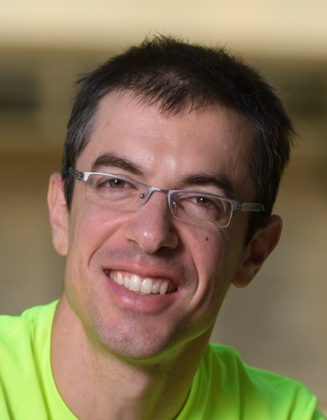
Dr. Yaron Orenstein : Deep learning for protein-RNA interactions (5/12/2018)

Protein-RNA binding, mediated through both RNA sequence and structure, plays a vital role in many cellular processes, including neuro-degenerative diseases. Modeling the sequence and structure binding preferences of an RNA-binding protein is a key computational challenge. Accurate models will enable prediction of new interactions and a better understanding of the binding mechanism.


In this talk, I will describe a new deep learning based approach to learn RNA sequence and structure binding preferences from large biological datasets. I will present the results of our algorithm outperforming the state of the art, both in vitro and in vivo. I will give examples of the biological insights we can gain by applying our neural networks to larger datasets of protein-RNA interactions. I will conclude with open questions and a discussion on the success of deep learning in computational biology.


No biological background is assumed or required for the purpose of the talk.

Bio:
Yaron is a Senior Lecturer at the Department of Electrical and Computer Engineering, BGU. He spent his post-doctoral training at CSAIL, MIT and the Simons Institute, UC Berkeley. Previously, he received his Ph.D and M.Sc degrees from CS and EE, TAU, respectively. Yaron completed his bachelor’s degree in Computer Science and Electrical Engineering at TAU.

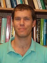
Oren Solomon: Fast Super-resolution Imaging in Optics and Ultrasound: From Sparsity to Deep Learning (21/11/2018)

Until recent years, the spatial resolution of diffractive imaging devices such as microscopes and ultrasound machines, was considered to be fundamentally limited, as first established by Ernst Karl Abbe almost 150 years ago. The 2014 Nobel prize in chemistry was awarded for methods which proved that although the diffraction limit poses a physical limitation, it can nonetheless be circumvented by altering the conventional measurement process in fluorescence microscopy. Drawing inspiration from microscopy, similar methods were applied to ultrasound imaging, achieving a precise mapping of sub-diffraction vascular networks deep within the tissue. However, although such techniques demonstrated unprecedented resolving power beyond the limit of diffraction, they lack in temporal resolution. Thus, the ability to image dynamic processes in sub-diffraction resolution is severely limited in these techniques.

In my work, I outline the main limitations of the pioneering super-resolution techniques, and present how fast super-resolution can be achieved by increasing fluorophore density and exploiting structural and statistical priors of the acquired signal. The first part of my work demonstrates that by exploiting sparsity in the correlation

domain, fluorescence microscopy can achieve sub-diffraction imaging with resolution comparable to state-of-the-art, while requiring two orders less the number of exposures. Next, I present how similar ideas can be extended to contrast enhanced ultrasound to achieve time-lapse imaging of super-resolved hemodynamic changes. Moreover, I also explain how in ultrasound we can further exploit the inherent motion of contrast agents to achieve Doppler processing in sub-diffraction resolution on one

hand, and on the other, how blood flow can be used as a structural prior for super-resolution. Lastly, I show that recent developments in the field of deep learning can also be applied to ultrasound imaging to achieve super-resolution, and to suppress tissue clutter signal for better visualization of blood vessels, as an initial step for further advanced processing.

BIO:

Oren Solomon is a Ph.D student under the supervision of Prof. Yonina Eldar.

Oren received his B. Sc. in electrical engineering from Ben-Gurion University , Beer-Sheva, in 2008 and his M. Sc. in electrical engineering from Tel-Aviv University in 2014. He is currently pursuing his Ph. D. degree in electrical engineering at the Technion-Israel Institute of Technology. His main research interests include theoretical aspects of signal processing, sampling theory, compressed sensing, medical imaging and optics, as well as deep learning. Oren was awarded The Andrew and Erna Finci Viterbi Fellowship Program for 2017.

Dr. Tamir Bendory: Estimation in extreme noise levels with application to cryo-electron microscopy (14/11/2018)

Single-particle cryo-electron microscopy (cryo-EM) is an innovative technology for elucidating structures of biological molecules at atomic-scale resolution. In a cryo-EM experiment, tomographic projections of a molecule, taken at unknown viewing directions, are embedded in highly noisy images at unknown locations. The cryo-EM problem is to estimate the 3-D structure of a molecule from these noisy images.

Inspired by cryo-EM, the talk will focus on two estimation problems: multi-reference alignment and blind deconvolution. These problems abstract away much of the intricacies of cryo-EM, while retaining some of its essential features. In multi-reference alignment, we aim to estimate a signal from its noisy, rotated observations. While the rotations and the signal are unknown, the goal is only to estimate the signal. In the blind deconvolution problem, the goal is to estimate a signal from its convolution with an unknown, sparse signal in the presence of noise. Focusing on the low SNR regime, I will propose the method of moments as a computationally efficient estimation framework for both problems and will introduce its properties. In particular, I will show that the method of moments allows estimating the sought signal accurately in any noise level, provided sufficiently many observations are collected, with only one pass over the data. I will then argue that the same principles carry through to cryo-EM, show examples, and draw potential implications.